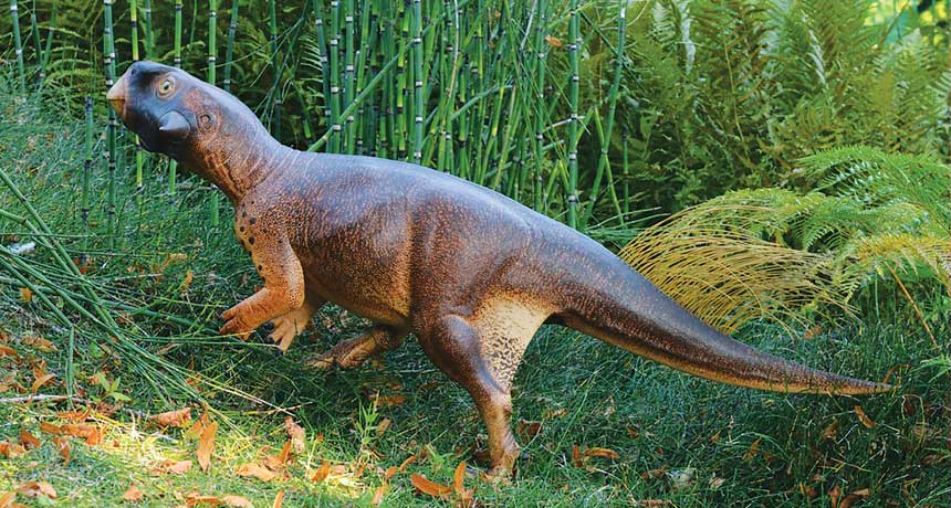
Dinosaurs may have used color as camouflage

The stories of dinosaurs’ lives may be written in fossilized pigments, but scientists are still wrangling over how to read them.
In September, paleontologists deduced a dinosaur’s habitat from remnants of melanosomes, pigment structures in the skin. Psittacosaurus, a speckled dinosaur about the size of a golden retriever, had a camouflaging pattern that may have helped it hide in forests, Jakob Vinther and colleagues say.
The dinosaur “was very much on the bottom of the food chain,” says Vinther, of the University of Bristol in England. “It needed to be inconspicuous.”
Identifying ancient pigments can open up a wide new world of dinosaur biology and answer all sorts of lifestyle questions, says zoologist Hannah Rowland of the University of Cambridge. “You might be able to take a fossil … and infer a dinosaur’s life history just from its pigment patterns,” she says. “That’s the most exciting thing.”
Not so fast, says paleontologist Mary Schweitzer of North Carolina State University in Raleigh. Evidence for ancient pigments can be ambiguous. In some cases, microscopic structures that appear to be melanosomes may actually be microbes, she says. “Both hypotheses remain viable until one is shot down with data.” Until then, she says, inferring dinosaur lifestyles from alleged ancient pigments is impossible.
Vinther’s work, published in the Sept. 26 Current Biology, is the latest in a long-simmering debate in the field of paleo color, the study of fossil pigments and what they can reveal about ancient animals. Disputes over his team’s findings and what’s needed to clearly identify fossilized melanosomes point to current pitfalls of the field.
But the promise is clear: Paleo color could paint a vivid picture of a dinosaur’s life, offering clues about behavior, habitat and evolution.
“This is a crucial new piece in the puzzle of how the past looked,” Vinther says.
Color me dino
Psittacosaurus (model shown) was a parrot-beaked herbivore about the size of a large dog. Researchers found signs of pigmentation (black specks) on its tail region, back leg and elsewhere that hint at its habitat.
Tap the image below to see signs of pigmentation from Psittacosaurus fossils.
A field emerges
Scientists have been puzzling over animals of the past for centuries, but eight years ago, paleontology got a wake-up call. That’s when Vinther and colleagues proposed that microscopic structures in a roughly 125-million-year-old fossil feather were actually a type of melanosome (SN: 8/2/08, p. 10). These pigment pouches rest inside pigment cells and, in this particular fossil feather, might have delivered a blackish hue, like a blackbird’s.
Scientists had noticed similar structures inside fossilized skin and feathers since the early 1980s. But people assumed that these structures were remnants of bacteria — perhaps decomposers that feasted on the dead animals, says paleontologist Martin Sander of the University of Bonn in Germany.
The new, colorful interpretation sparked a flurry of research, and scientists have since spotted what appear to be melanosomes in all kinds of fossilized animals. Paleontology, in fact, is now awash in colors and patterns. Pigment pods may have painted reddish-brown speckles on the face of a Late Jurassic theropod, brushed chestnut stripes on a long-tailed dino from China and made the plumage of a four-winged dinosaur called Microraptor iridescent. That shimmery dinosaur “probably had a weak, glossy iridescence all over its body,” says evolutionary biologist Matthew Shawkey of Ghent University in Belgium. His team deduced Microraptor’s color from the shape of its melanosomes.
Modern melanosomes generally carry a mixture of two melanin pigments: dark brown-black eumelanin and red-yellow pheomelanin. Scientists have linked color in mammals and birds to melanosome shape — a meatball shape for reddish brown hues, for example, and a sausage shape for darker colors.
In iridescent feathers, melanosomes tend to be even thinner, Shawkey says. Microraptor’s melanosomes looked like skinny sausages — similar to those seen in the feathers of modern crows and ravens, says Shawkey, who reported the findings with Vinther and colleagues in Science in 2012 (SN Online: 3/9/12).
Three years later, Vinther laid out the case for inferring color — and ancient histories — from fossilized pigments in a review in Bioessays. Not only can the distinctive shapes of melanosomes offer clues, he noted, but chemical tests can help detect the presence of melanin itself. Finding this pigment in fossils, he argued, puts the old bacteria hypothesis to rest.
Schweitzer and colleagues disagreed with Vinther’s take in a review published in Bioessays later in 2015. Researchers need to be cautious when deducing the hues of extinct animals, the scientists wrote. Any melanosome look-alikes in fossilized feathers or skin could actually be microbes.
After all, microbes are everywhere. “These animals died in an environment that was not sterile and free from microbes,” Schweitzer says. “Think about it. If you take a piece of chicken and throw it out in your backyard, how long does it take for microbes to overgrow that chicken?”
The tiny organisms are hardy, too. Both microbes and the sticky biofilms they form are preserved in the fossil record. And, Schweitzer says, microbes and melanosomes overlap completely in shape and size, which makes the two tough to tell apart. What’s more, some microbes actually make melanin themselves; detecting the pigment in a fossil is not a rock-solid sign that the ancient animal was black, brown or freckled.
It’s not that Schweitzer or Bioessays coauthor Johan Lindgren, a geologist at Lund University in Sweden, doubt that melanosomes can leave traces in the fossil record. The issue, Lindgren says, is that not all round structures you find are melanosomes.
Chemical tests could help distinguish the two. Bacteria, for example, leave behind traces that can be identified with pyrolysis gas chromatography-mass spectrometry. But that requires samples to be vaporized. “It can mean destroying much of what you are trying to study,” says geochemist Roy Wogelius of the University of Manchester in England. “So it’s not always possible.”
Vinther’s new work isn’t likely to settle the debate. In fact, people were arguing both sides in October at a meeting of the Society of Vertebrate Paleontology in Salt Lake City.
Arindam Roy, a Bristol colleague of Vinther’s, reported size differences between fossilized melanosomes and bacteria growing on decaying chicken feathers in the lab. Alison Moyer, an N.C. State colleague of Schweitzer’s, said that looks weren’t enough. Finding keratin, a protein that typically surrounds melanosomes, could serve as evidence for pigments in fossils.
From color to camouflage
The fossil described in Vinther’s new paper is “spectacular,” Schweitzer says. “It’s got skin all over the place. I can’t think of too many dinosaur specimens that are preserved like this.”
The dinosaur lies on its back, flattened in a slab of volcanic rock. Skin covers a completely intact skeleton, and dozens of long bristles poke from the tail. Psittacosaurus, an herbivore that lived some 120 million years ago, walked on two legs and would have reached about half a meter in height.
“It would have been a supercute animal,” Vinther says. “It’s got this wide face and looks a little bit like E.T.”
Black material speckles the dinosaur’s body, tail and face. Vinther believes the material is the ancient remains of pigment. His team examined samples chipped from the fossil and saw what he considers the telltale orbs of melanosomes — mostly impressions in the rock but also some microbodies, the 3-D structures themselves.
Based on the dinosaur’s pigment patterns, it would have had a dark back that faded to a lighter belly. That type of coloring, called countershading, shows up in animals from penguins to fish and may act as a form of camouflage. It lightens parts of the body typically in shadow, and darkens parts typically exposed to light. “If you want to hide, it makes sense to try and obliterate those shadows,” Rowland says.
Their prediction for diffuse light matched the model painted like Psittacosaurus. “It’s like what we see in forest-living animals,” Vinther says. “This thing was camouflaged.”
Lingering doubts
Going from fossil to forest may be more of a leap than a step, other scientists suggest.
Psittacosaurus’ skin very well may contain ancient pigments, Wogelius says. “I don’t think it’s a crazy idea.” But, he adds, of Vinther’s group: “I don’t think they’ve proved what they claim.”
Vinther’s team, for exampl e, used just four tiny fossil samples to extrapolate the coloring of the whole dinosaur. “I think it’s a bit of an overreach,” Wogelius says.
Schweitzer also notes that the specimen was varnished, presumably to protect the bones and soft tissues. It happened before Vinther and colleagues got their hands on the dinosaur and makes it impossible to perform the chemical tests that would bolster the claim for pigments. “Varnish is horribly destructive to fossils,” she says. “It totally ruins the specimen for other types of analysis.”
Vinther argues that his team has chemically analyzed other fossils and found evidence of melanin — not bacteria. The microbodies in those fossils look just like the ones in Psittacosaurus, he says.
Vinther’s team also saw evidence of just one kind of microbody, and it had a distinct round shape. If the structures were actually bacteria, he says, you’d expect to see a whole range of shapes and sizes. “Some of them would be shaped like corkscrews, some would have flagella, some would be humongous, some would be tiny.”
That’s the tricky part with bacteria, counters Lindgren. “In some cases you can have a huge consortium, but in other cases you can have one single type.”
Vinther’s interpretation has its supporters. “I was skeptical at first,” Sander says, “but now there’s been such an array of these little bodies that it’s pretty clear that at least some of them are not bacteria.” Despite some continuing controversy, Sander says many paleontologists now accept that microstructures in fossils may be melanosomes.
Additional research, though, “would help the entire community,” he says, “so that there are no longer any lingering doubts.”
Along with chemical tests, Schweitzer suggests, researchers could try transmission electron microscopy, a technique that blasts an electron beam through a thinly sliced sample. With TEM, melanosomes appear as black blobs. Bacteria tend to look different — in some cases, more like fried eggs.
Shawkey, for one, is looking to chemistry. In a paper published online November 14 in Palaeontology, his team used a technique called Raman spectroscopy to help build a case for feather color in a bird that died some 120 million years ago. In the feathers, the researchers spotted the skinny sausages of iridescent melanosomes and chemical signs of the pigment eumelanin. Shawkey thinks the chemical evidence could help “head off any criticism that we might encounter.”
Working through the field’s snags, paleontologists might come together to fill in the hues and tints, and potentially the habits and habitats, of ancient animals that until recently had been known primarily by their bones.